


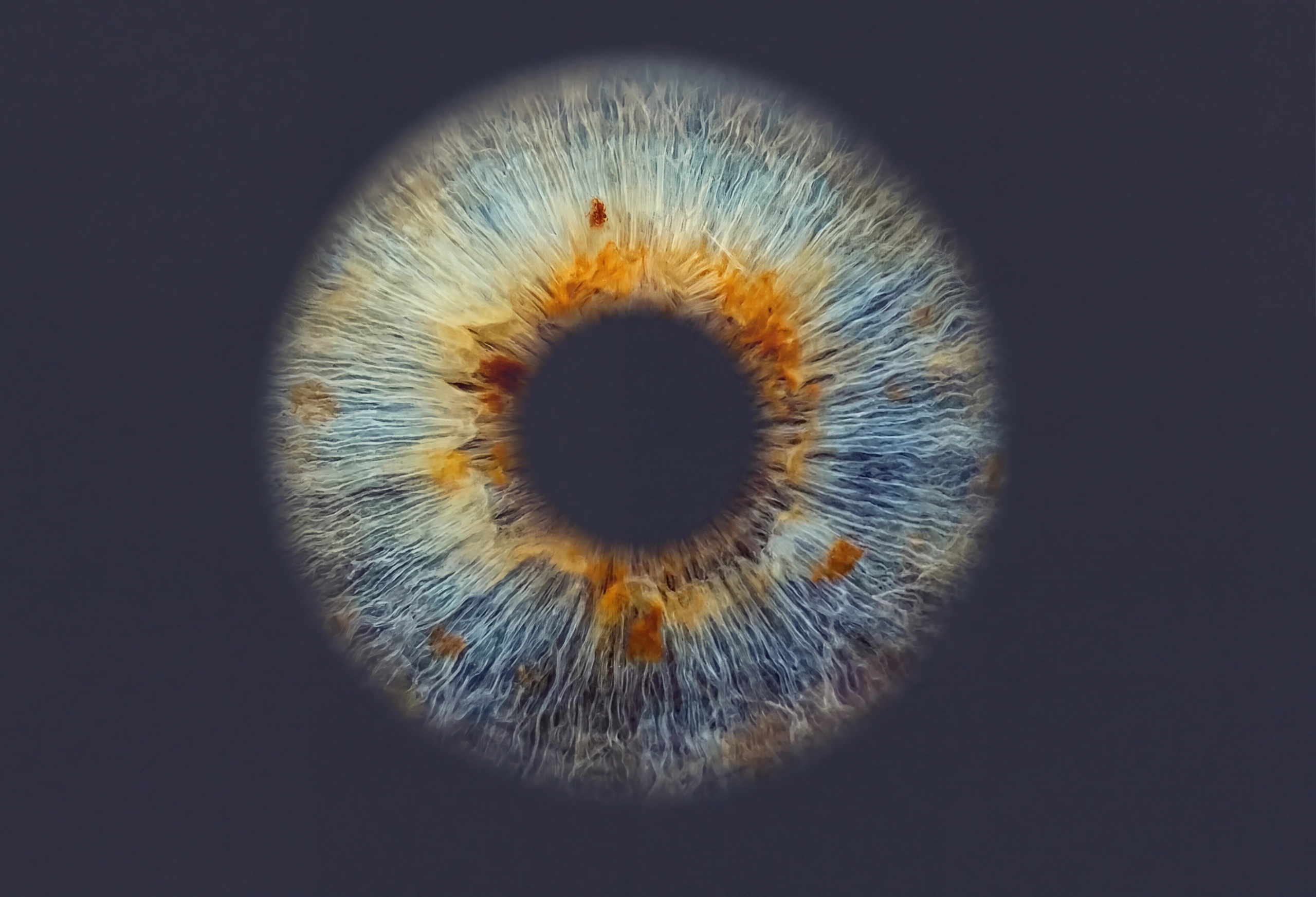
Chromatic Pupillometry for Photoreceptor Function Analyses
retinaWISE is an advanced binocular chromatic pupillometer that assesses pupil dynamics under spectrally controlled light stimuli. It enables objective detection of retinal functional changes, with the goal to support early assessment and treatment-efficacy follow-up.
- Multiple light sources to selectively stimulate photoreceptor classes, combined with binocular pupil dynamic measurements.
- Dichoptic light paths to deliver stimuli that are independent of pupil size.
- Pre-configured and custom protocols for clinical assessment and research.
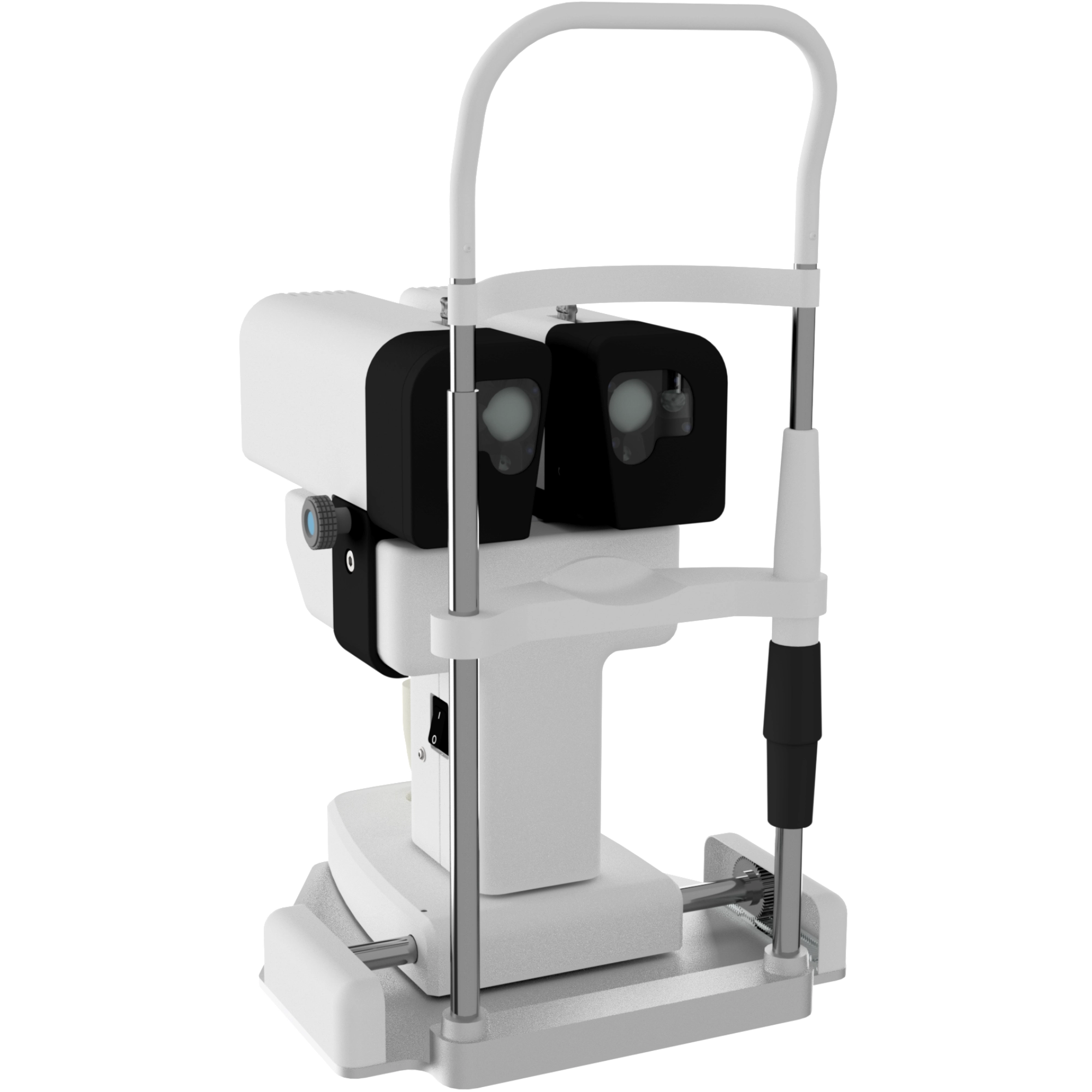
Bilateral Tracking of Pupil Response to Light Stimuli
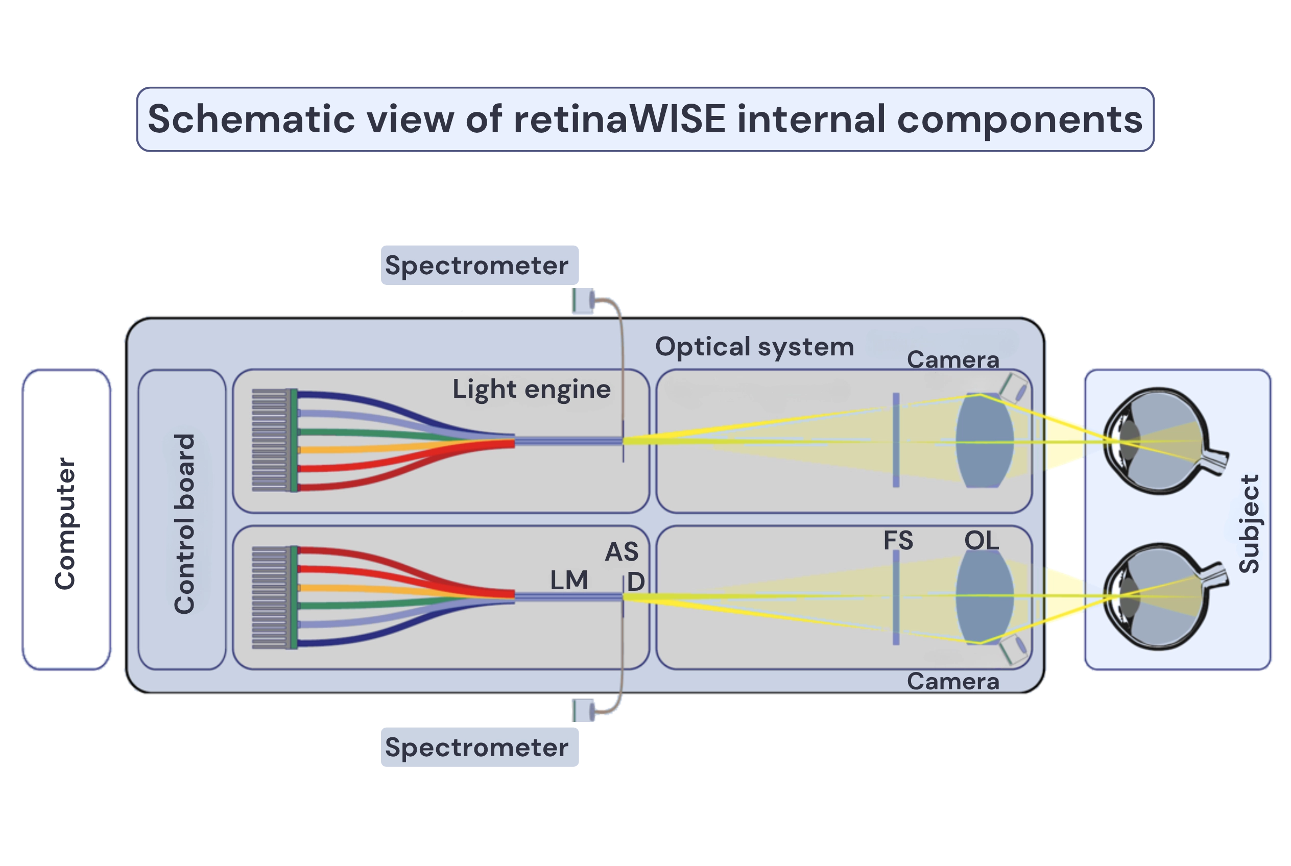
Six monochromatic LEDs, Maxwellian optical arrangement, and bilateral independent pupillometry combined to make measurements objective, reproducible, and specific to the targeted photoreceptor class.
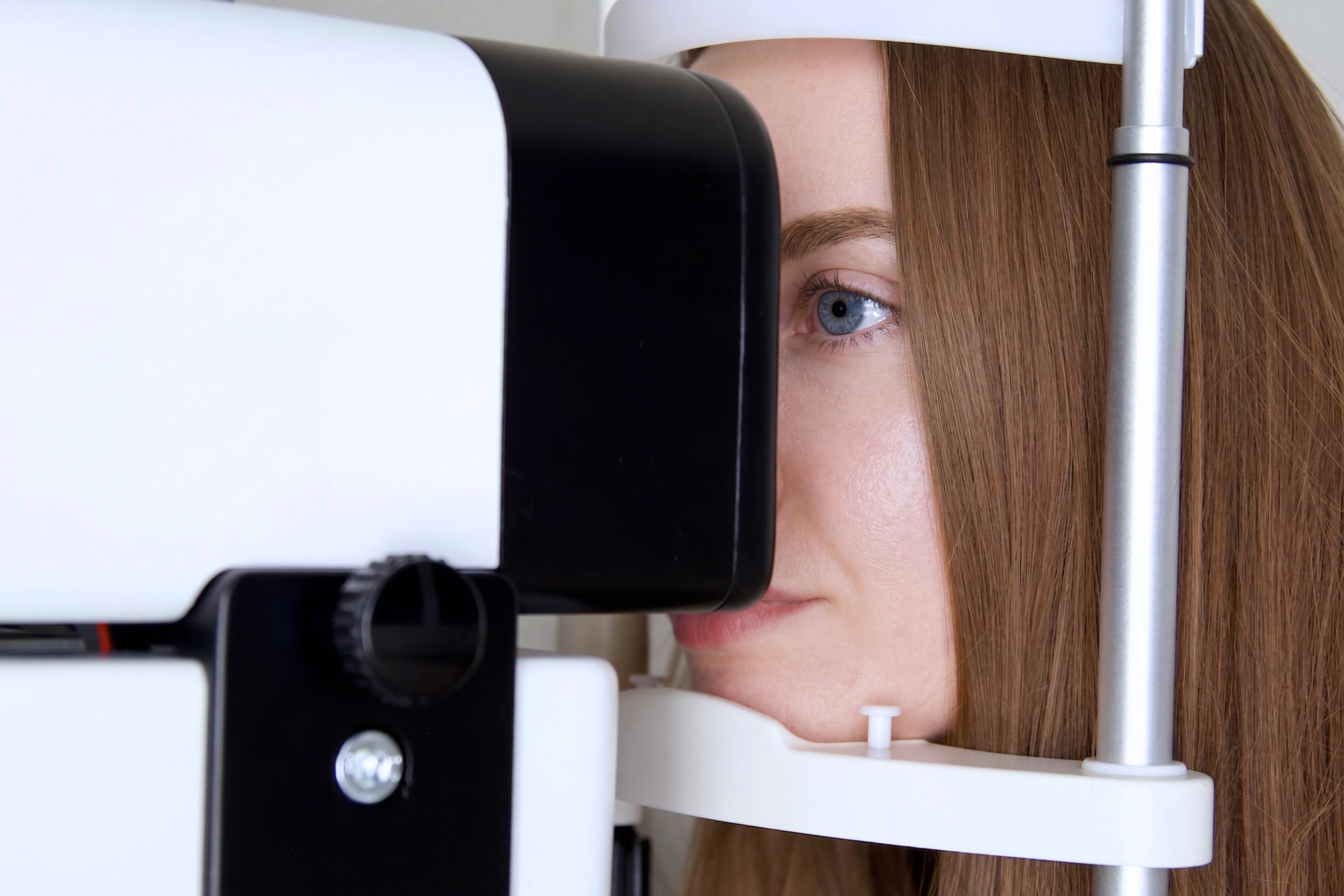
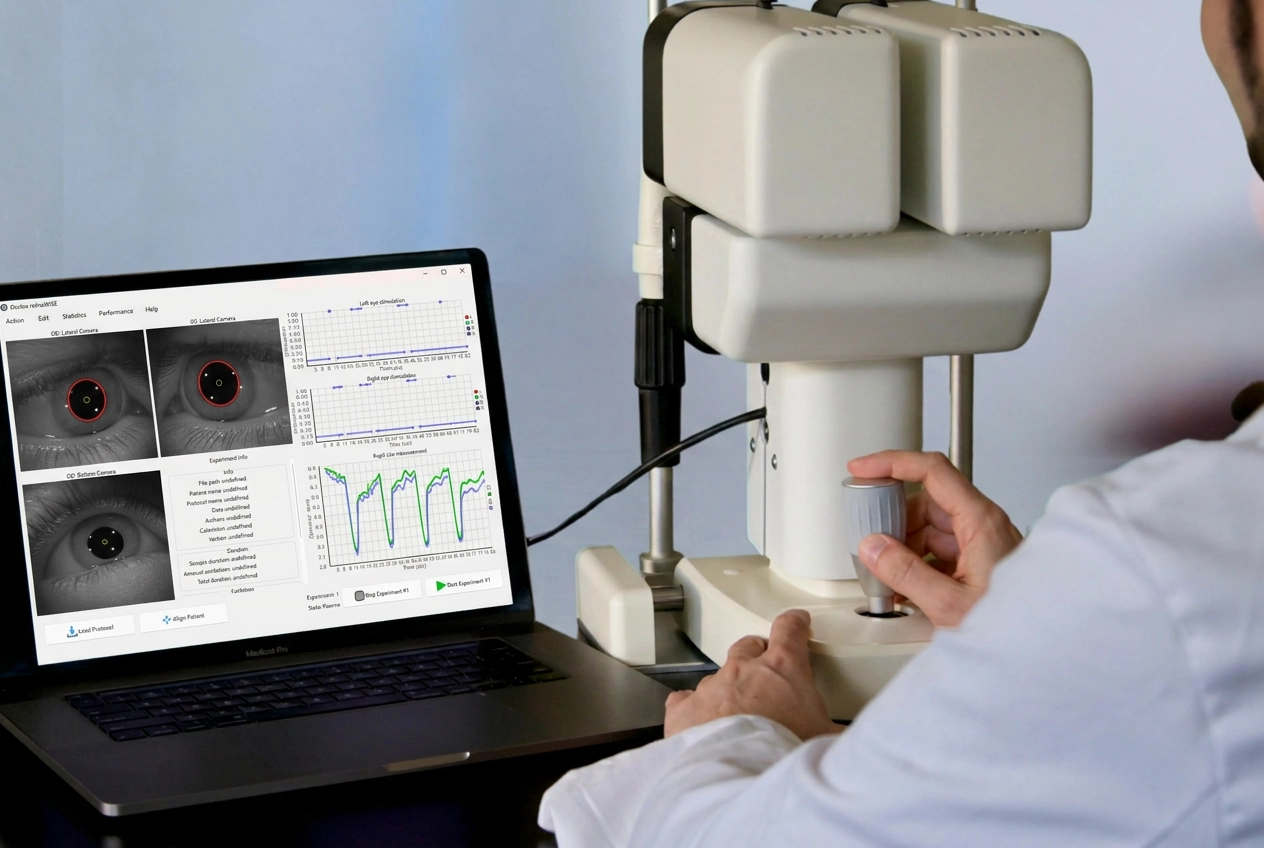
Patient Alignment & Setup
Light Stimulus Configuration
Bilateral Stimulation & Measurement
Data Collection & Analysis
What retinaWISE Measures
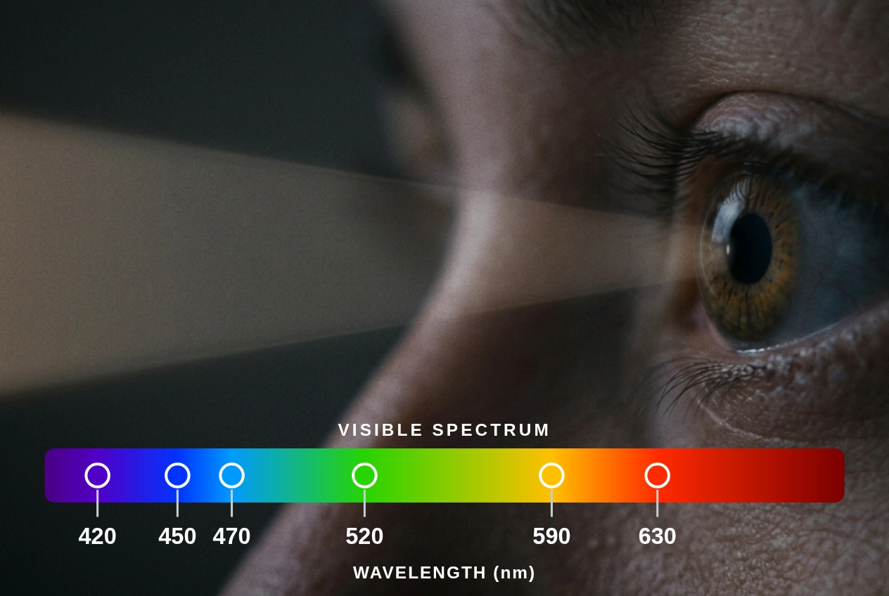
retinaWISE measures the pupillary light reflex (PLR) under spectrally controlled light stimuli to assess the function of rods, S-cones, M-cones, L-cones, and ipRGCs1. Photoreceptor- and melanopsin-mediated pupil responses have been studied as objective markers of retinal and optic-nerve function in different clinical conditions2 including AMD3, glaucoma4-5, and neurodegenerative disease6.
Organisation of the human retina: incident light traverses the inner retinal layers to reach the outer segments of photoreceptors adjacent to the retinal pigment epithelium (RPE), where phototransduction occurs. In parallel, intrinsically photosensitive retinal ganglion cells (ipRGCs), containing melanopsin, are involved in pupillary light reflex regulation and circadian rhythm entrainment.7
Rod Photoreceptors
Scotopic (low-light) VisionHighly light-sensitive rods mediate dim-light vision. Peak spectral sensitivity ~498 nm. Isolated with low-intensity or scotopic stimuli followed by a dark adaptation time.
Rod-mediated pupillary responses have the lowest threshold of activation. Low-irradiance blue light can be used to test rod function in outer retinal diseases.2
Cone Photoreceptors
Photopic & Colour VisionS-, M- and L-cones mediate daylight and colour vision. Peak sensitivities ~420, 530 and 560 nm. Each class is isolated via silent substitution across the 420–630 nm wavelength range.
Cone-mediated responses help assess maculopathies and cone dystrophies.2
ipRGCs
Non-visual & Circadian RhythmIntrinsically photosensitive retinal ganglion cells (ipRGCs) contain melanopsin and act as non-visual light sensors impacting circadian rhythms. Peak sensitivity ~480 nm. Stimulated with high-contrast blue light via silent substitution.
Mediated by ipRGCs, the sustained post-illumination pupil response (PIPR) can be altered in AMD,3 glaucoma,4-5 and Alzheimer's disease.6
Intended Use & Opportunities
retinaWISE measures pupillary dynamics in response to selective chromatic stimulation of photoreceptors (rods, S-, M- and L-cones) and ipRGCs. This device could support early functional assessment and treatment follow-up across ocular and neurological disorders. The areas below indicate active fields of clinical and research application. Disease-specific validation is ongoing.
Research Collaborations

Our research groups adopted retinaWISE as part of two research programmes: HELIOS-BD and AMBIENT-BD, which investigate light sensitivity and monitor circadian rhythms in bipolar disorder. retinaWISE enables selective, independent stimulation of the photoreceptors involved in circadian regulation.

retinaWISE is implemented at the Centre for Sleep and Circadian Science to assess mood disorder risk through the post-illumination pupil response (PIPR), as part of research on sleep and circadian rhythms in adolescents and young adults.

The Centre for Chronobiology at the University Psychiatric Clinics (UPK) Basel employs retinaWISE to measure the effects of caffeine on light processing in adolescents and adults. retinaWISE allows for selective stimulation of specific photoreceptors to isolate melanopsin contributions.
Research Behind the Technology
The development of retinaWISE, and research studies employing it, are described in peer-reviewed publications.
Diagnostics (Basel) · MDPI · PubMed-indexed
Development of an Innovative Pupillometer Able to Selectively Stimulate the Eye's Fundus Photoreceptor Cells
Peer-reviewed description and characterisation of the retinaWISE instrumentation, demonstrating selective photoreceptor stimulation via silent substitution and a Maxwellian optical arrangement.
View publicationWellcome Open Research
HELIOS-BD: Non-Visual Responses to Light in Bipolar Disorder
Clinical-research protocol applying chromatic pupillometry with retinaWISE to assess melanopsin-pathway and non-visual responses to light in bipolar disorder.
View publicationGet Started with retinaWISE
Clinicians: Request device specifications or arrange a live demonstration of retinaWISE.
Researchers: Enquire about research site access, protocol customisation, or existing study data.
Institutions: Contact Oculox to discuss evaluation agreements or distribution.
retinaWISE: CE-marked Class I medical device, certified November 2023 under EU Medical Device Regulation 2017/745.
For objective photoreceptor function assessment.
Work Behind Chromatic Pupillometry
Chromatic pupillometry, including assessment of the Post-Illumination Pupil Response (PIPR), is described in peer-reviewed literature as a method for evaluating photoreceptor and melanopsin (ipRGC) function across retinal, optic nerve, and neurological conditions:
1. Gibertoni G, Hromov A, Piffaretti F, Geiser MH. Development of an Innovative Pupillometer Able to Selectively Stimulate the Eye’s Fundus Photoreceptor Cells. Diagnostics 2024;14:1940. DOI: 10.3390/diagnostics14171940
2. Rukmini AV, Milea D, Gooley JJ. Chromatic pupillometry methods for assessing photoreceptor health in retinal and optic nerve diseases. Front Neurol. 2019;10:76. DOI: 10.3389/fneur.2019.00076
3. Maynard ML, Zele AJ, Feigl B. Melanopsin-mediated post-illumination pupil response in early age-related macular degeneration. Invest Ophthalmol Vis Sci. 2015;56(11):6906–6913. DOI: 10.1167/iovs.15-17357
4. Najjar RP, Sharma S, Aung T, Milea D, et al. Pupillary Responses to Full-Field Chromatic Stimuli Are Reduced in Patients with Early-Stage Primary Open-Angle Glaucoma. Ophthalmology. 2018;125:1362. DOI: 10.1016/j.ophtha.2018.02.024
5. Kelbsch C, Maeda F, Strasser T, et al. Pupillary responses driven by ipRGCs and classical photoreceptors are impaired in glaucoma. Graefes Arch Clin Exp Ophthalmol. 2016;254:1361. DOI: 10.1007/s00417-016-3351-9
6. La Morgia C, Mitolo M, Romagnoli M, et al. Multimodal investigation of melanopsin retinal ganglion cells in Alzheimer's disease. Ann Clin Transl Neurol. 2023;10(6):918–932. DOI: 10.1002/acn3.51773
7. Masland RH. The neuronal organization of the retina. Neuron. 2012;76(2):266–280. DOI: 10.1016/j.neuron.2012.10.002